Sample Case Descriptions
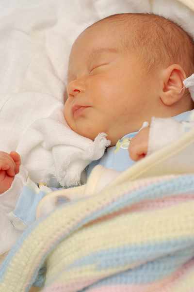
A term infant was born via C-section, due to failure to descend and failed trial of forceps, resulting in sustained permanent brain injury from acute, near total asphyxia. Neurologically speaking, neonatal depression was noted with evidence of posturing and possible seizure activity on the day of the birth, at which point the infant was started on phenobarbital. I independently examined the child at four years of age and rendered an opinion on causation.
Infant was born five days prior to the estimated date of confinement. Medications during pregnancy included Cervidil, Pitocin, Cleocin and Ambien. Complications during the pregnancy and delivery included maternal fever to near 102. Medications during labor and delivery included Cleocin. Membranes had been noted to be ruptured for 18 hours and the amniotic fluid was meconium stained. Patient was born by vaginal delivery, with hypoxic ischemic insult and traumatic injury to the brain due to vacuum extraction. Birth weight was 3.775 kg and Apgar scores were 0 at 1 minute, 0 at 5 minutes, and 1 at 10 minutes. From a neurological standpoint, the infant had "perinatal depression", signs of neonatal encephalopathy on the first day of life, seizure activity shortly after birth and an EEG was consistent with encephalopathy with multifocal sharp waves in both hemispheres as well as seizures originating in the left temporal and right central regions. The baby was eventually discharged on phenobarbital. I provided a causation opinion.
A term infant girl was born by emergency cesarean section with alleged delay in her delivery. APGAR scores were 1 at 1 minute and 3 at 5 minutes. She was initially diagnosed with hypoxic ischemic encephalopathy and respiratory failure. Within hours of being born she was noted to have seizures. MRI imaging of the brain showed signs of acute injury to the basal ganglia region. She was ultimately diagnosed with quadriparetic cerebral palsy. I was asked to provide my opinion as to the cause of the child’s hypoxic ischemic encephalopathy and discuss the extent of her injury.
17 year old girl presented to the emergency room with a history of acute onset decreased mental status and trouble speaking. She was initially diagnosed as having a psychiatric illness and psychiatry was consulted. She had normal labs and a normal CT scan of the brain at admission. She was observed over several hours while awaiting transfer to a psychiatric hospital. Eight hours after presenting to the ER she was noted to have right face, arm and leg weakness. She was sent back for further neuroimaging and was found to have had a middle cerebral artery stroke. I was asked to provide my opinion as to the cause of the stroke and the likelihood of any treatment being effective in reducing the damages this stroke caused.
A term infant male was born to an obese, diabetic mother. Shoulder dystocia was diagnosed during the delivery. The initial examination of the child after being born showed weakness of the entire arm with normal finger movements. The child was ultimately diagnosed with an upper brachial plexus injury (Erb’s Palsy) that was permanent. I was asked to provide testimony on the causation of a brachial plexus injury and her physical limitations.
The mother of a preterm (34 week) infant female was involved in a severe automobile collision. Within one hour of the injury the child was born by spontaneous vaginal delivery. Shortly after being delivered she had neonatal encephalopathy and seizures. Initial abdominal imaging showed a liver hemorrhage and imaging of the brain showed subarachnoid and subdural hemorrhages. The child was alleged to have developmental and cognitive delays secondary to this injury. I was asked to provide causation testimony.

Pediatric Neurology Expert, Brian E. Woodruff MD